Products

Meet FIM.

Field Induced Mydriasis — the world's first device to dilate the pupil without drops, without contact, and without the wait.

90–120s
Time to full dilation
<120s
Recovery to normal condition
The clinical term

What is Mydriasis?

Mydriasis is the medical term for pupil dilation. FIM stands for Field Induced Mydriasis — inducing this exact clinical state without drugs, drops, or contact. The visuals below show how a dilated pupil (mydriasis) compares to a normal, constricted (miosis), or unevenly sized (anisocoria) pupil.
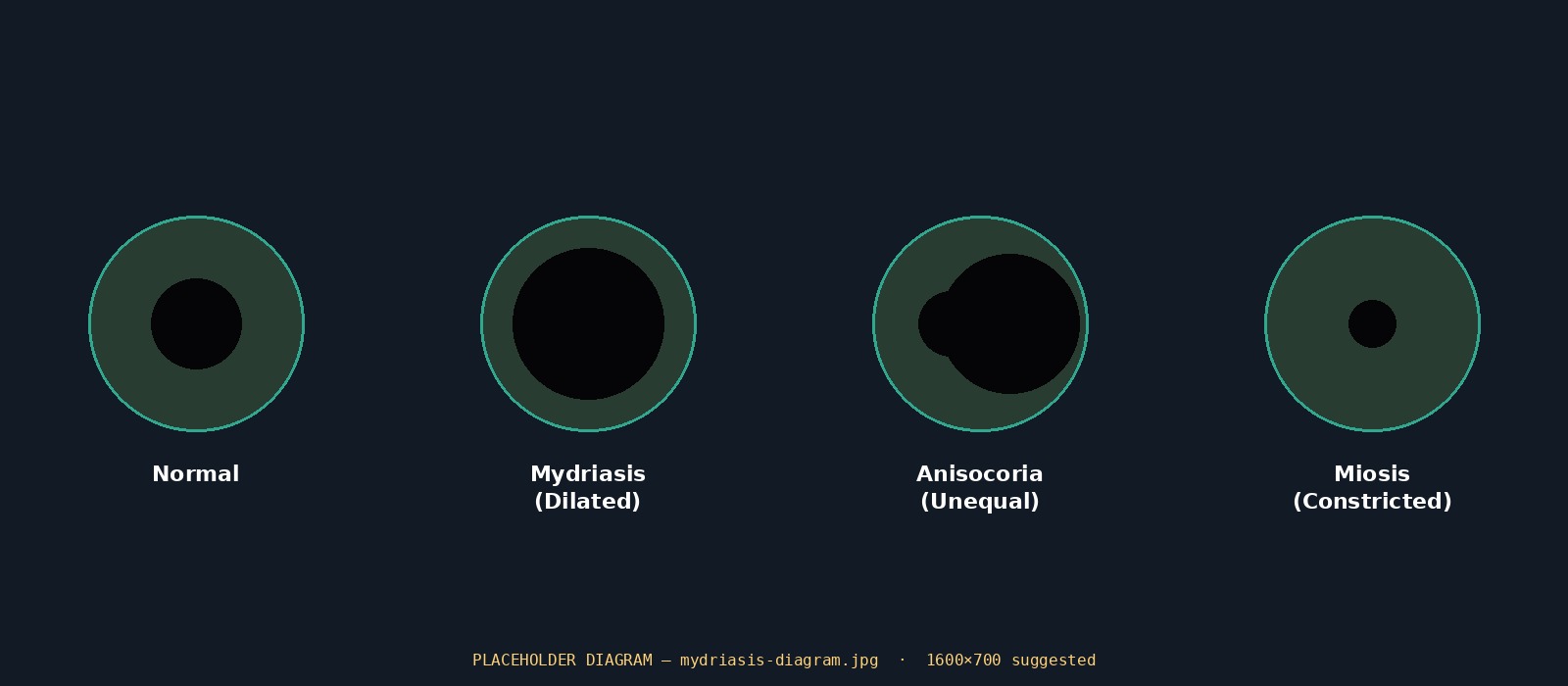
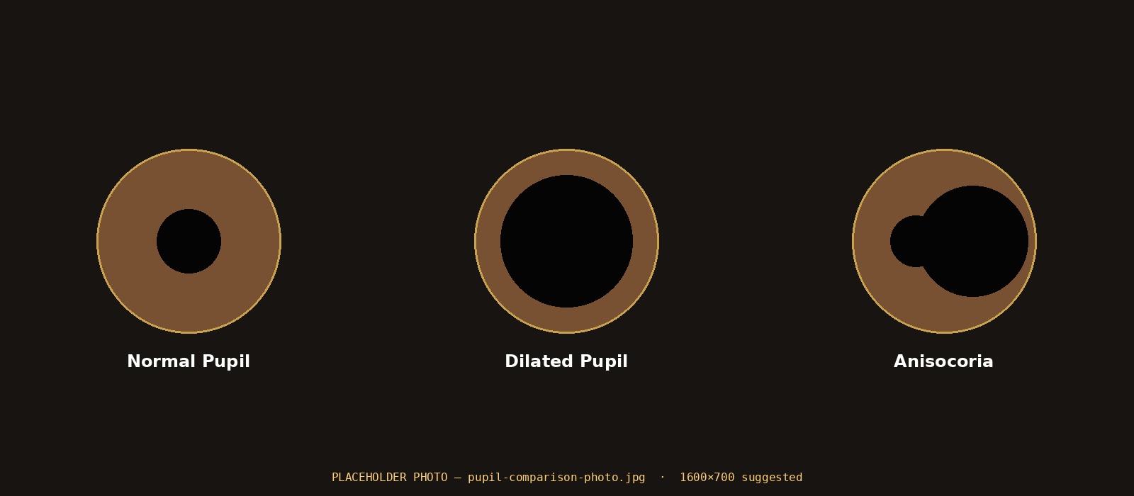
Diagram comparing normal pupil, mydriasis, anisocoria and miosis
Real eye photos comparing normal pupils, dilated pupils and anisocoria
Real Time Pupil Dilation

See FIM in action

Captured by a leading digital imaging system using proprietary ATI software for precise measurement of pupil size, under normal eye examination room conditions.

How it works

Drops out. Field in.

FIM is easily installed on any standard slit lamp. Its operation is seamlessly integrated with the standard ophthalmology examination procedure without pre-examination preparations or waiting periods.

1. Patient is seated

The patient sits in front of the slit lamp, as in a regular procedure, placing their head within the headrest.

2. Doctor adjusts the scope

The doctor adjusts the slit lamp microscope for the examination, exactly as in a regular procedure.

3. FIM switches on

The doctor turns FIM on and waits up to two minutes, until the pupil is fully dilated.

4. Exam proceeds

The OFE is adjusted to maintain ~12–15mm from the patient's eye, and the doctor proceeds with the exam.

5. FIM switches off

At the end of the procedure, the doctor simply turns FIM off, or the patient can remove their head from the chin rest.

6. Fast recovery

The pupil recovers to normal condition within minutes, without any side effects.

System components

What's in the box

Ocular Front-end Unit (OFE)

Generates the safe electromagnetic field that induces pupil dilation. Mounted on the slit lamp in front of the examined eye at ~12–15mm, with a hollow ring aligned to the slit lamp's microscope. Optional lenses can be inserted as in regular exams.

FIM Ocular Front-end Unit
FIM Generator Box Unit

Generator Box Unit (GBU)

Holds the system software, LED signals and controls, generating all signals converted into electromagnetic fields at the OFE. Simple on/off switch with LED feedback lights, powered via a standard medical power adapter.

Why it matters

Eye drops vs. FIM

Traditional eye drops

  • Requires contact with the eye
  • 15–30 minutes to reach full dilation
  • Blurred vision for hours afterward
  • Not suitable for some patients

FIM

  • Non-contact, drug-free, no medication
  • 90–120 seconds to full dilation
  • Recovery to normal condition in minutes
  • No pain or discomfort reported
Rocket launch — FIM 2nd generation is headed to space
What's Next

FIM 2nd Generation is headed to space

ATI is developing the next generation of FIM — and one of its future versions is planned to fly on a space mission, extending the technology beyond the clinic and into a new frontier for ophthalmic diagnostics.

Read the Full Story
Earth seen from the lunar surface
Beyond orbit

A new vantage point for diagnostics

Exploration on a planetary surface
Built for extremes

Eye care wherever the mission goes

Certifications

Approved and audited

July 2014CE Marking of Conformity renewed
July 2014ISO 13485:2003 certificate renewed
November 2013Israel Ministry of Health AMR Marking of Conformity
October 2012CE Marking of Conformity received
October 2012ISO 13485:2003 certificate received
Specifications

The details

Time to full dilation90–120 seconds
Recovery time<120 seconds
Contact with eyeNone
Medication requiredNone
CertificationCE, ISO 13485:2003, Israel MOH AMR
InstallationFits any standard slit lamp
Patient discomfortNone reported

Seamless workflow

Integrates into standard slit lamp examination procedures with no added steps for staff.

Safe by design

Uses safe electromagnetic fields conforming to CE standards — no medication, no needles, no drops.

NIT — personal ocular tonometer
In development — NIT

Glaucoma detection, in patients' hands

Ocular hypertension is the most important risk factor for glaucoma, but today's gold-standard tonometer can only be used by a professional. ATI is developing NIT, a personal ocular tonometer offering fully automatic IOP measurement — enabling self-examination by patients at home.

The non-invasive measurement is performed on a closed eye via the eyelid, using image processing of a reference pattern that deforms under applied pressure — making frequent, timely glaucoma screening accessible outside the clinic.

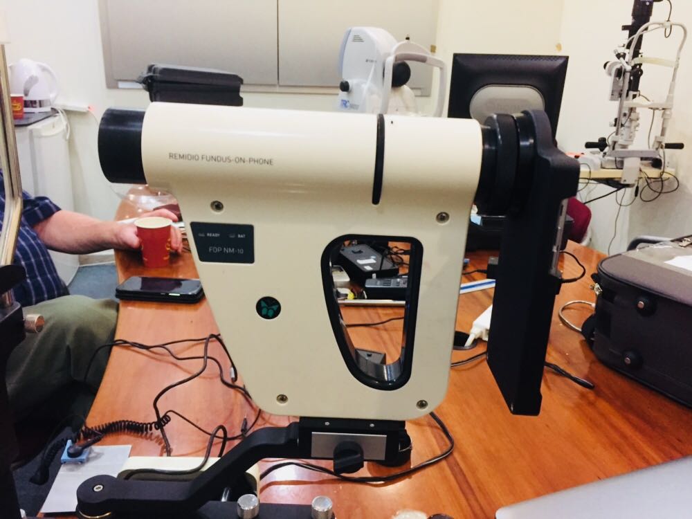
FIM integration with Remidio Mobile Fundus Camera
Ask us
anything
Get FIM in your clinic

Ready to see it for yourself?

We're happy to walk you through a live demo, share clinical data, or talk through what it takes to get FIM installed at your practice.

Request a Demo